However, many experts emphasise educational pressures are not the only cause. The rapid growth of cities, as well as a decline in traditional close-knit family structures, have also been attributed to increased feelings of isolation, depression and suicide.
This content is available customized for our international audience. Would you like to view this in our US edition?
Sometimes, when the weather closes in, physicians have to be creative, said Shirk. "I’ve done an appendectomy with both Dr. Cullen and with Dr. Todd; those are surgeries that can happen here with caution."
Retrobulbar optic neuritis is an inflammation of the optic nerve posterior to the optic nerve head. As a result, anterior and posterior segment evaluation of the eye is unremarkable, as was the case with our patient. In addition, patients will complain of eye pain that worsens with eye movements. However, this condition was ruled out due to a lack of an afferent pupillary defect, no reduction in visual acuity and normal nerve appearance with MRI.

Posterior scleritis is also inflammation of the sclera, but it occurs posterior to the ora serrata. Presentation varies, but signs can include optic nerve swelling, retinal hemorrhages, choroidal folds, hyperopic shift, exudative retinal detachment, choroidal detachment, restricted motilities and proptosis. The patient may come in complaining of eye pain worse with movement, double vision or blurred vision. Imaging is imperative in the diagnosis of posterior scleritis. B-scan ultrasonography can reveal subretinal fluid in Tenon’s capsule and posterior scleral wall thickness greater than 2 mm in approximately half of patients (Lavric, et al.). These findings are considered pathognomonic for posterior scleritis. Ultrasound, OCT and MRI or CT will show thickening of the sclera and/or choroid. Although our patient did present with severe eye pain, none of the posterior segment findings were present, and there was no choroidal thickening on MRI of the orbits.
Orbital pseudotumor, also known as idiopathic orbital inflammatory syndrome, is a nonspecific inflammation of orbital tissue. Patients with orbital pseudotumor will present with periorbital pain, eyelid erythema and swelling. Depending on the severity, swelling and inflammation may result in proptosis and ophthalmoplegia. MRI or CT scan will reveal orbital inflammation. Proptosis, ophthalmoplegia, erythema and swelling of the eyelids were all absent in our patient. MRI ruled out orbital pseudotumor due to the lack of orbital inflammation.
Scleritis is inflammation of the sclera, the white fibrous tissue underneath the conjunctiva and episclera, and its blood vessels. It predominantly occurs in women, and its incidence increases with age. Scleritis can be idiopathic but is often related to underlying autoimmune disease. It is characterized by deep boring eye pain and presents as a red eye that does not blanch with instillation of 2.5% phenylephrine (Gerstenblith and Rabinowitz). Episodes of scleritis can result in thinner areas of sclera, presenting as a gray or blue patch of sclera. Gray patches of sclera were present in our patient’s left eye. However, active scleritis was easily ruled out due to the white and quiet appearance of the patient’s sclera and no intraocular inflammation. The gray patch of the sclera in our patient was deemed to most likely be scleral melanocytosis, a congenital gray-blue pigmentation of sclera not associated with systemic conditions. Oculodermal melanocytosis, also known as Nevus of Ota, involves the sclera and eyelids and is associated with an increased risk of uveal melanoma.
I wrote mildly homoerotic manic street preachers fan fiction for a GCSE creative writing assignment. It was deemed unsuitable.

Differential diagnoses for our patient included: scleritis, retrobulbar optic neuritis, orbital myositis and orbital pseudotumor. These etiologies were ruled out with a careful case history, ocular evaluation and appropriate imaging. (Of note: MRI with contrast and fat suppression is preferred when evaluating soft tissue.)
The PVT is a small plastic scope that attaches to your phone (check the product page for compatibility) and runs each eye through a series of simple app-powered tests.
In some sinusitis cases, more severe complications can occur if a bacterial superinfection develops. Although rare, inflammation or infection of certain sinuses – namely the sphenoid, ethmoid and frontal sinuses – carries a higher risk of secondary complications. Because of their anatomic locations, sphenoidal and ethmoid sinusitis are the most likely to lead to cavernous sinus thrombosis. Orbital cellulitis is more likely to arise from ethmoid sinusitis because of the paper-thin bony wall (lamina papyracea) that separates the ethmoid sinus from the orbit. Infection in the frontal sinus has an increased risk of spreading to the brain, causing meningitis or encephalitis. For this reason, MRI is indicated in patients with suspected complications of acute sinusitis who present with complications lasting longer than 48 hours after initiation of treatment.
(Editor’s note: Family physicians provide 42 percent of care(www.ruralhealthweb.org) in rural communities. AAFP News will highlight some of these FPs over the next few months. The series will focus on why some family docs choose to practice in small towns off the beaten path and how they provide high-quality, comprehensive care to patients of all ages — often with far fewer resources than their urban and suburban colleagues.)
PHOTOS. Blessé, Neymar s’éclate au Brésil pendant le carnaval de Rio | Pillow Block Related Video:
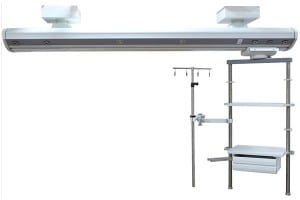
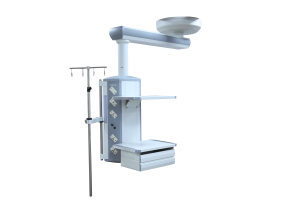
We take "customer-friendly, quality-oriented, integrative, innovative" as objectives. "Truth and honesty" is our administration ideal for Medical Operation Table, Theatre Light, Led Medical Examination Lamps, We have been fully aware of our customer's needs. We deliver high quality products, competitive prices and the first class service. We would like to establish good business relationships as well as friendship with you in the near future.

